Echocardiogram (Echo)

  • Home / Department / Echocardiogram (Echo)

Echocardiogram (Echo) is a graphic outline of your heart’s movement. During this test, high-frequency sound waves, called ultrasound, provide pictures of your heart’s valves and chambers. This allows your doctor to see the pumping action of the heart.

To schedule a Echocardiogram (Echo) Test, please contact us today.

Doctors

Our Specialist

Internist/Nephrologist Dr. Sheyla Zelaya and Internist Dr. Ana Karovska have built a new model for their practice on the ‘qualities’ that allow the best medical practices to succeed and flourish - getting close to thier patients, close to their needs, their concerns, their hopes and their dreams.

Sheyla Zelaya, MD

Nephrology/Internal Medicine

Ana K. Vuchidolov, MD

Board Certified - Internal Medicine

Frequently asked questions

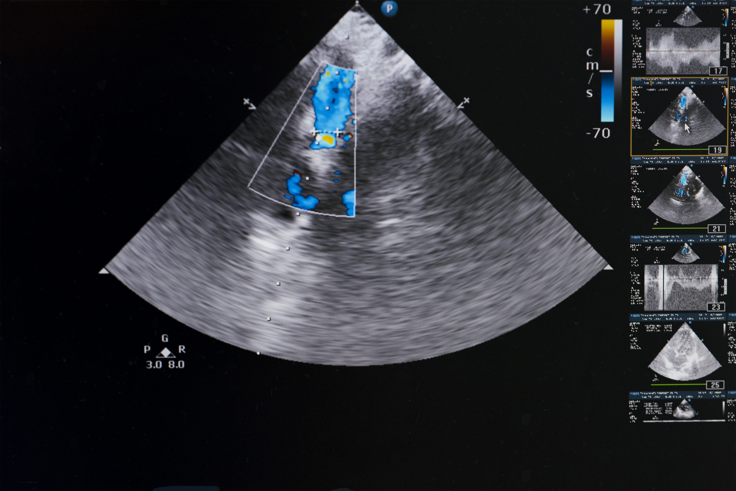
An echocardiogram may be suggested by your heart doctor if he or she suspects complications within the chambers or valves of your heart. Typically, after doing a physical examination and learning more about your symptoms and health background, your cardiologist may have a good idea what tools need to be used in order to diagnose the problem. This particular form of testing is often used to measure pumping function in those with heart failure; it may also be used to determine the extent of damage to the heart after a heart attack. An echocardiogram may be helpful in evaluating, diagnosing and monitoring many conditions, including:

  • Abnormal heart valves
  • Atrial fibrillation
  • Congenital heart disease
  • Heart murmurs
  • Infections in the sac around the heart
  • Pulmonary hypertension
  • Transthoracic echocardiogram (TTE) – This is the most common, standard, non-invasive echocardiogram. In this test, a transducer is placed firmly against the outside of your chest to pick up sound waves, which are then converted into moving images on a screen.
  • Stress echocardiogram – This type of echocardiogram monitors and takes ultrasound images of your heart activity before and after performing physical activity (such as walking on a treadmill).
  • Doppler echocardiogram – This test is performed in order to monitor speed and direction of blood flow through blood vessels, valves and heart chambers. This may help your doctor see if there are any irregularities or problems in your blood flow.
  • Transesophageal echocardiogram (TEE) – If the picture on the transthoracic (regular) echocardiogram is unclear (sometimes due to lung disease or obesity), a TEE may provide a much clearer picture. For this procedure, a tube is guided down the esophagus, so that it can be placed very close to the heart for an unobstructed view. This test is performed under mild sedation.
  • Transthoracic echocardiogram – There is little to no risk involved.
  • Transesophageal echocardiogram – It’s common to have some throat soreness after the procedure is finished. This should improve within hours of the procedure.
  • Stress echocardiogram – Typically after performing exercise, your heart rate is elevated and may be irregular for a short time. More serious complications, such as a heart attack, are very rare.

Putting Quality into Practice

It’s an honor and a privilege to participate in your care. As you get to know us, we believe you will see
how important it is for us to get to know YOU. We believe g ood communication is a vital skill for
everything from understanding your concerns to explaining a diagnosis, to relaying information across
the healthcare system to other healthcare professionals who may be involved in your care.