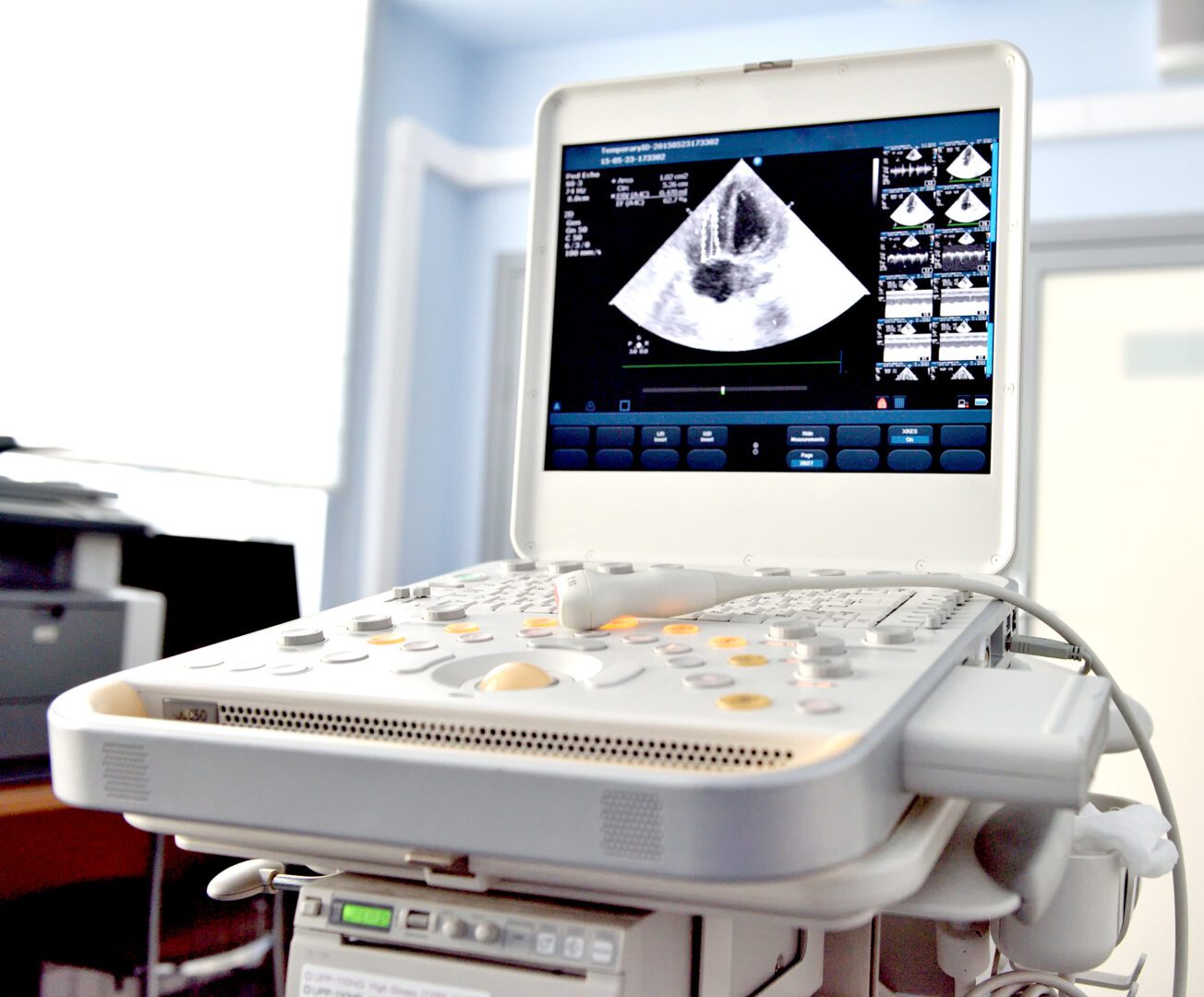
The ultrasound scanner, also known as a sonogram, is the most versatile imaging tool. It allows radiologists to examine nearly every organ in the body.
Ultrasound uses high-frequency sound waves to produce a visual of internal organs and structures of the body. A transducer, or wand or probe, produces these sound waves that travel through tissues and fluids.
The sounds waves bounce back off denser tissues and structures; this is known as an echo. The denser the object is the greater the echo. The returning sounds waves produce the images in real-time, which means that ultrasound imaging can show the movement of the organs and blood flow.
Freequently asked questions
Ultrasound scanners use a wand-like transducer to produce sound waves. These waves travel harmlessly through the body, reflecting and bouncing off organs. These sound waves return to the transducer, producing a real-time image visible on a computer monitor.
Radiologists routinely use ultrasounds for numerous purposes. We use it to evaluate the thyroid, to look inside the gallbladder for gallstones, to diagnose appendicitis and to examine pelvic and abdominal organs. They also help radiologists detect masses, blood clots and narrowing in the arteries that could lead to a stroke. Ultrasounds are also widely used on pregnant women to monitor an unborn child’s development and to check for birth defects.
In most cases, ultrasound exams do not require preparation. Depending on the area that needs to be scanned, you may need to fast or drink water in advance. During the exam, the technologist will apply a thin layer of water-gel to your skin or on the transducer. The gel aids in the transmission of the sound waves through your body. The technologist will place the transducer on your skin and move it around to get an accurate picture. Depending on the type of exam you may need, the transducers could be used internally, such as during a vaginal ultrasound. When the scan is over, the technologist will wipe the gel off from your skin and you can return to your normal activities. Most ultrasounds generally take around 20 to 30 minutes to complete.


